Jochen Weiss, DJ McClements and P Michael Davidson
Introduction
Globally, approximately 2.2 million people are killed annually because of foodborne and waterborne diseases with numbers continuing to rise due to increase in resistance of pathogens and emergence of new pathogens (Food Standards Agency UK 2011). An example of the latter is the outbreak of a novel strain of Escherichia coli O104:H4 in Germany in May of 2011. The outbreak led to 48 deaths, 857 hemolytic uremic syndrome (HUS) cases and 3078 non-HUS cases (WHO 2011). Due to the high import – export trade volume of Germany, the outbreak quickly spread to other countries with which Germany was doing business at the time.
An old but increasingly revisited solution to this problem is the addition of antimicrobials to foods (Davidson et al. 2005). In contrast to antibiotics, only temporary and no genetic resistance developments take place when using these substances which is the reason why even after humankind having used them for thousands of years they still continue to function. Antimicrobials have been defined as substances that inhibit the growth and/or inactivate microorganisms. Historical records indicate that the Babylonians, ancient Greek and Romans already used vinegar to preserve food, and that nitrates were apparently used as early as in Homer’s time (850 BC) to preserve meat. However, many antimicrobials suffer from high losses of activity when used in our increasingly complex modern food products (Weiss et al. 2011). Using novel nanotechnological approaches to make them work better thereby improving safety and shelf life of food products has recently been seen as a way to overcome this problem.
Nanotechnology, i.e. the application of nanoscale sciences, has generally gained considerable attraction in various industrial fields, chief amongst them the chemical, pharmaceutical, automotive and electronics industries (Weiss et al. 2007). Materials that are structured and engineered at the nanoscale have been shown to possess unique attributes since quantum mechanical effects rather than Newtonian physics govern physical properties. Electromagnetic, and not gravitational forces dominate, and the importance of particle-wave matter duality and tunneling effects increase. Key physicochemical characteristics such as melting point, boiling point, conductivity and solubility become size dependent on the nanoscale. The latter, that is solubility, often increases as size decreases, a fact that is particularly relevant for the bioactivity of many molecules. Thus, nanostructuring of bioactives is being seen as a mean to improve their efficacies (Chen et al. 2013). It is particularly this fact that has caused the food and pharmaceutical industries to become interested in nanosciences. Since most bioactive materials do not form nanostructures on their own, carrier structures providing the desired nanoscalar properties must be designed and manufactured (Weiss et al. 2009). Considering the above mentioned issues of new pathogens and resistance developments, the interest in the use of nanocarriers to potentially increase activity of antimicrobials is understandable.
In this article, we will provide a brief overview over nanocarrier systems that have been used to carry and deliver antimicrobials. Showing some select results from recent studies, we will highlight that choosing appropriate structures can indeed lead to activity increases, but that conversely an inappropriate structure design can also lead to activity losses. We will also discuss some issues surrounding the incorporation of such structures in complex food matrices and finally highlight alternative approaches to improve the activity of antimicrobials in foods.
Encapsulation of Antimicrobials in Nanocarriers
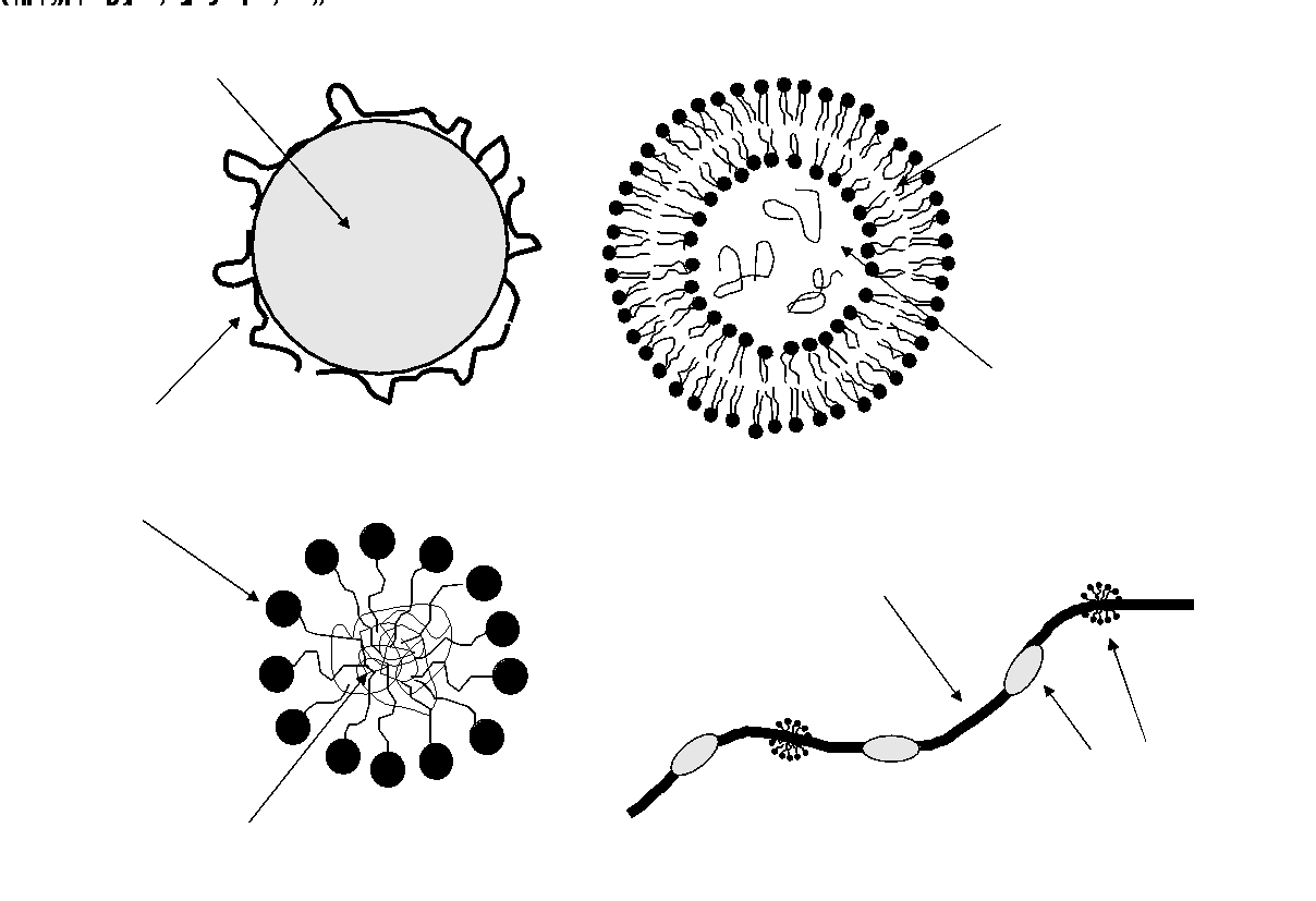
In recent years, a wide variety of different nanoscalar encapsulation systems have been developed for use in the food and pharmaceutical industries (Weiss et al. 2009). It is virtually impossible to provide a full review of them considering that studies describing the design of new structures are published almost on a daily basis. Interested readers may want to consult a number of excellent recent reviews on this topic (McClements 2012, 2013). Below we will instead provide a brief overview of four specific nanoscalar encapsulation systems that have shown particular promise in delivering antimicrobials in foods, namely nanoemulsions, liposomes, microemulsions and nanofibers. Figure 1 provides a brief schematic overview over these four systems and illustrates which components in these different structures could possess antimicrobial properties.
Figure 1. Schematic illustration of four nanoscalar encapsulation systems: (top left): oil-in-water emulsion; (bottom left) microemulsion; (top right) liposomes; (bottom right) electrospun nanofibers. The figure also illustrates which components may possess antimicrobial activity.
Antimicrobial nanoemulsions
Emulsions are systems that are composed of two or more completely or partially immiscible liquids with one liquid being dispersed in the other in the form of droplets (Weiss et al. 2007, McClements 2012, Weiss et al. 2000) . Emulsions can be manufactured by a wide variety of different techniques such as high pressure homogenisation, membrane extrusion, microfluidics, sonication or phase inversion (Yang et al. 2012). Depending on the method used, they can be made to contain very fine disperse droplets, e.g. droplets have mean droplet diameters ranging between 50 to 150 nm. In such cases, they are referred to as nanoemulsions. As a recent in-depth review pointed out, such nanoemulsions differ from conventional emulsions in a number of ways (McClements 2013). For example, they can be transparent or nearly translucent since droplets are so small that they do not scatter light in the visible range. The small particle size also leads to very low rates of gravitational separation. This becomes quickly evident when considering the Stokes equation, which states that the velocity of a particle moving in a gravitational field due to density differences is proportional to the square of the diameter of the particle. Thus reducing the particle diameter by a factor of two leads to a decrease in the Stokes velocity by a factor of 4. On the other hand, nanoemulsions must contain substantially more emulsifier than normal emulsions. Emulsifiers are amphiphilic, surface active that during emulsion manufacturing adsorb to droplet interfaces thereby lowering interfacial tension and providing repulsive interacting forces that prevent droplets fromaggregating or coalescing. Since nanoemulsions have much larger interfacial areas than conventional emulsions at similar oil droplet concentrations, much more emulsifier is needed, which can be a regulatory or organoleptic issue.
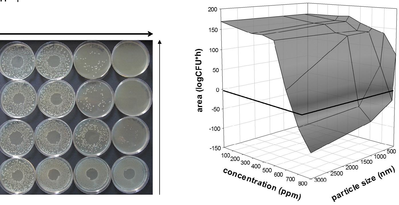
Emulsions and nanoemulsions have been used in recent years as carrier systems for hydrophobic antimicrobials such as for example certain essential oil components (carvacrol, eugenol, thymol) or parabenes, and for amphiphilic antimicrobials such as lauric arginate, nisin or lysozyme (Donsi et al. 2011, Ferreira et al. 2010, Karthikeyan et al. 2011, Sznitowska et al. 2002, Terjung et al. 2012). Activity studies of these systems against foodborne pathogens or spoilage organisms however have had many researchers puzzled, since nanoemulsions were sometimes more active and sometimes less active than macroemulsions. This is also illustrated in Figure 2 that shows results of a model pathogen (E. coli C 600) growth test where cultures were incubated with emulsions carrying eugenol as an antimicrobial (Terjung et al. 2012). The emulsions had different mean oil droplet, ranging from approximately 3 nm to 80 nm. The photographic images of the plates illustrate that the antimicrobial emulsions were quite active and gradually killed E. coli cells over time. After 24 and 48 h the impact of the different mean droplet sizes of emulsions became clearly visible. Surprisingly, it was the emulsions with larger droplet sizes that were more effective over time than the smaller ones. A quantification of this effect done by integrating the growth curves as a function of time to calculate a net effect further confirmed these results (right hand side of Figure 2). There, large negative values indicate that a substantial net kill occurred. Conversely, large positive values indicate that cells grew. Net effect values were smaller and became even positive when emulsion droplets became smaller, indicating that the emulsions became less effective. This is in contrast to other studies with bioactive compounds that had indicated that bioactivity may be enhanced when encapsulating them in nanoemulsions (Donsi et al. 2011, Karthikeyan et al. 2011, Jain et al. 2011).
Figure 2. Results of an antimicrobial activity test using E. coli C 600 as target microorganisms. (Left) Photographic images of plates incubated for various times containing 800 ppm eugenol that had been incorporated in emulsions having particle sizes from 80 to 3000 nm (Right) Net effect of antimicrobial emulsion treatment expressed as area obtained by integration of the growth curve a period of 24 hours. Results are shown as a function of particle size of the emulsion and concentration of eugenol in the emulsion (Terjung et al. 2012)
We have since come to understand that this is because very complex distribution and interaction phenomena with microorganisms, the solvent phase and the oil phase of the emulsion take place. In some case, making the emulsion smaller improves their interaction with the target microorganisms, increases diffusional mass transport leading to a more homogeneous distribution of the antimicrobials throughout the entire food system. In other cases though ̶ especially if antimicrobials are strongly bound to droplet interfaces and droplets don’t interact directly with microbial cells ̶ antimicrobials may no longer be released from oil droplets and therefore fail to inhibit pathogens or food spoilage organisms. The example above therefore illustrates that nanoscaling does not always lead to the desired outcome.
Antimicrobial microemulsions
Amphiphilic molecules composed of hydrophilic and hydrophobic groups dispersed in solvents can spontaneously self-organise to form micelles, bilayers and reversed micelles (Weiss et al. 2007, Weiss and McClements 2000).The aggregates have well-defined properties, e.g. size and charge. The process occurs spontaneously and is physically rather than chemically driven. Organic compounds of low solubility can be brought into solution by addition of surfactants (Weiss et al. 2000, Gaysinksy et al. 2005, Weiss et al. 1996, 1997). The increase in solubility in the presence of surfactants is known as solubilisation. McBain and Hutchinson (Sjoeblom 1967) defined solubilisation as ‘a particular mode of bringing into solution substances that are otherwise insoluble in a given medium, involving the presence of a colloidal solution whose particles take up and incorporate within or upon themselves the otherwise insoluble material.’ Solubilisation has been of interest to scientists in various fields of research such as cosmetics, pharmaceuticals, insecticides, herbicides and detergents (Sjoeblom 1967, Tadros 1980, Carroll et al. 1982). It is also one of the most frequently used reasons why surfactants are employed, since solubilisation is the basis for detergency.
The ability of micelles to act as carriers for compounds with limited water solubility has been used to improve the delivery of antimicrobials (Yang et al. 2012, Gaysinksy et al. 2005, Fu et al. 2006). Studies have focused on solubilising essential oil compounds such as carvacrol (2-methoyl-5-[1-methylethyl]phenol) and eugenol (2-methoxy-4-[2-propenyl]-phenol) in surfactant micelles to produce so-called “microemulsions” with the purpose of increasing the activity of encapsulated antimicrobials (Gaysinksy et al. 2005a,b). Aqueous solutions of Surfynol® 485W and 465, two model nonionic surfactants, prepared at a range of concentrations (1 - 10% w/w) were able to solubilise two potent essential oil components, namely eugenol and carvacrol. These antimicrobial microemulsions were transparent, stable at various pH and temperatures and effective against a range of bacterial pathogens including E. coli O 157:H7 and Listeria monocytogenes.
Antimicrobial liposomes
Liposomes are spherical bilayer structures composed of polar lipids (Taylor at al. 2005). Industrially, they are most often produced by high pressure homogenisation similar to the procedure most often used for the manufacturing of emulsions. The homogenising process involves passing a mixture of active ingredient, water and phospholipids though a valve causing spherical liposomes to be generated (Hofland et al. 1993). Depending on the applied pressure in the homogeniser, liposomes may contain only a single bilayer membrane, in which case the are referred to as small (< 100 nm) or large (> 100 nm) unilamellar vesicles, or SUVs and LUVs, respectively (New 1990). If the mixture above is not passed through an homogeniser and is instead only mixed such that low shear stresses are applied, then liposomes containing more than a single bilayer membrane may be formed. These are known as multilamellar vesicles (MLV, if all layers are concentric), or multivesicular vesicles (MVV, if a number of randomly sized vesicles is enclosed in the interior of another vesicle) (New 1990).
Liposomes can serve as a separate microenvironment in which active components can be incorporated and their activity be maintained despite changes in the surrounding aqueous phase (Bolsman et al. 1988, Bouwstra 1996). Thapon and Brule (1986) used liposomes to encapsulate lysozyme and nisin to prevent spoilage of cheeses. Degnan and Luchansky (1992) reported anti-listerial effects of the bacteriocin, pediocin AcH, in beef tallow and muscle slurries using PC liposomes. Bacteriocin activity increased by 28% following encapsulation compared to non-encapsulated pediocin. Authors reported that pediocin AcH activity decreased rapidly when unencapsulated bacteriocin was added to slurries or tallow, likely a function of cross-reactions with food component leading to loss of activity (Degnan and Luchansky 1992). Benech et al. (2002a,b) encapsulated nisin Z (an asparagine-containing variant of nisin) in hydrogenated PC liposomes (1 mg nisin Z/mL buffer) and added liposomes or unencapsulated nisin Z to cheese at a final concentration of 300 IU/g. Listeria innocua counts decreased by 3 log CFU/g over a six-month ripening period (Benech et al. 2002a,b). Nisin Z activity in liposomes at the end of the six-month ripening period was ~90% of the initial activity, with a final concentration of L. innocua below 10 CFU/g cheese.
Tables 1 and 2 show select results of from our own studies in which nisin and lysozyme were incorporated in small unilamellar liposomes to yield antimicrobially active vesicles (Taylor et al. 2007a,b, Were et al. 2004a,b). The polar lipid composition of the liposomes was modulated by using mixtures of phosphatidylcholine (PC), phosphatidylglycerol (PG) and cholesterol. The liposomes were applied at similar active compound concentration against 5 different strains of L. monocytogenes. One can rapidly see another surprising result, namely that the two antimicrobials function quite differently in the liposomes. Nisin works much better when encapsulated, particularly in PC or PC:cholesterol liposomes, while it is just the opposite with lysozyme which works better in its free form. This is likely due to nisin and lysozyme interacting in a very different manner with the polar lipid which in turn may lead to the antimicrobial being either in the liposomal membrane or in the interior of the liposome. One can imagine that the protective effect of the liposome as well as the release behavior would then be quite different.
Table 1. Log reduction of various L. monocytogenes strains incubated for 30°h at 35°C in the presence of liposomes containing nisin
|
Strain |
PC |
PC:Cholesterol |
PC:PG:Cholesterol |
Unencapsulated |
|
101 |
>2.3 |
>2.3 |
0.8 |
1.3 |
|
108 |
>3.1 |
>3.7 |
0.7 |
1.0 |
|
310 |
>3.7 |
>3.7 |
0.7 |
0.8 |
|
V7 |
>2.8 |
>2.8 |
1.1 |
1.5 |
|
Scott A |
>2.6 |
0.5 |
1.4 |
1.9 |
Table 2. Log reduction of various L. monocytogenes strains incubated for 30°h at 35°C in the presence of liposomes containing lysozyme
|
Strain |
PC |
PC:Cholesterol |
PC:PG:Cholesterol |
Unencapsulated |
|
101 |
0.4 |
1.9 |
1.7 |
>2.3 |
|
108 |
0.5 |
0.3 |
0.4 |
>3.1 |
|
310 |
0.6 |
>2.6 |
>2.6 |
>2.6 |
|
V7 |
0.1 |
1.2 |
0.6 |
>1.7 |
|
Scott A |
< 0.1 |
1.3 |
>1.3 |
>1.3 |
Antimicrobial nanofibers
Electrospinning is a manufacturing technique that can be used to produce thin fibers from (bio-)polymer solutions with diameters below 100 nm (Kriegel et al. 2008). In electrospinning, polymer nanofibers may be obtained by application of a high voltage static electrical field that is formed between a grounded target and a polymer solution that is pumped from a storage chamber through a small capillary orifice. The high electric field causes a polymer solution jet to emerge from the tip of the capillary orifice. Since the jet contains charged polymer molecules, the jet velocity increases with increasing distance from the orifice and decreasing distance to the target causing the jet to become thinner and thinner. The thinning of the fiber facilitates a rapid evaporation of the solvent thereby inducing a transition from a liquid to a solid. The solid fibers are subsequently deposited on the grounded target and can be collected there as a non-woven mesh or membrane.
Electrospun nanofibers have been shown to possess a number of beneficial properties that distinguish them from larger nonwoven fibers produced by other methods. Aside from having higher tensile strengths than conventional fibers – a byproduct of the above described electrospinning process that forces polymer strands to strongly align – fibers have extremely high surface to volume ratios. This high surface area facilitates for example catalytic conversions or rapid release of small molecule compounds incorporated in the fibers. While initially only non-foodgrade electrospun fibers were manufactured due to the poor ability of water to act as an electrospinning agent, the later use of organic acids followed by crosslinking of biopolymers rendering them insoluble and thus washable, has yielded foodgrade fibers.
Electrospun nanofibers can be rendered antimicrobially active in a variety of ways. Firstly, the (bio-)polymers used as matrix material for the fibers may be an antimicrobial (Kriegel et al. 2009a). Figure 3 shows an example of such a system (unpublished data). There, chitosan, a cationic polysaccharide that exhibits strong antimicrobial activity, was used to fabricate fibers. On the left hand side of Figure 3 an image of chitosan fibers obtained by scanning electron microscopy can be seen. There, average fiber sizes are well below 100 nm demonstrating that the fibers were truly nanoscalar.
Figure 3. (Left) Scanning electron microscopy image of an electrospun chitosan nanofiber network at a resolution of 20.000. (Right) Cell numbers of a Salmonella spp. culture incubated for 100 h in the presence of various amounts of chitosan nanofibers
On the right hand side results of an antimicrobial effectiveness test of the fibers can be seen. To this purpose, fibers were suspended in a standard culture medium containing Salmonella spp. and cell counting was performed as a function of time. Cell counts decreased the higher the fiber level added to the system. The results suggest that there could be some advantages when using these types of systems as active packaging materials or as processing aids. Another possibility of rendering fibers antimicrobial is by including antimicrobially-loaded emulsion droplets or microemulsions in the polymer solution prior to the electrospinning process (Arecchi et al. 2010, Kriegel et al. 2009b, 2010). Studies have indicated that microemulsions and small emulsion droplets can then become an integral part of the fiber releasing their content at a later time when, for example, suspended in a solution.
Issues Surrounding the Use of Nanoscalar Dispersions of Antimicrobials in Complex Matrices
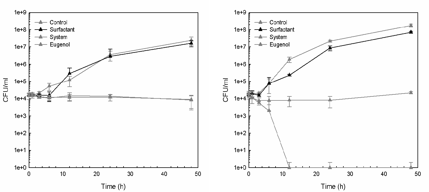
The discussion above shows that design and formation of nanoscalar dispersions of antimicrobials is not a trivial matter. Depending on the composition, the colloidal structure and the spatial arrangement of compounds in said structure, the system can attain quite unexpected functionalities. These could be both desirable and undesirable. To complicate the matter further, not only do these systems have unexpected functionalities in solution, they can also react in unforeseen ways with ingredients present in complex food structures. To illustrate this, results of an antimicrobial inactivation test conducted with the above mentioned microemulsions carrying eugenol as antimicrobial in either skim or whole milk are shown in Figure 4 (Gaysinsky et al. 2007). While the microemulsion with eugenol is able to rapidly inactivate L. monocytogenes cells in skim milk over the course of approximately 12 h, the same system is only able to keep cell numbers constant rather than decreasing them.
Figure 4. Results of an antimicrobial activity test in skim and while milk with antimicrobial microemulsions prepared from a 5 wt% aqueous surfactant solution (Surfynol® 485W) with 0.5 wt% eugenol against Listeria monocytogenes Scott A. (Left) while milk, (right) skim milk (from Gaysinsky et al. 2007)
Likely, the microemulsion in the whole milk system not only releases eugenol to the target microbial cells but also releases eugenol into milk fat droplets rendering the system less active. There are in fact many more examples like this that show that upon incorporation of such systems in complex food matrices activity may change. Loss factors though may vary greatly depending on the product in which the nanoscalar dispersion is applied. Microemulsions have for example been found to work quite well in apple juice and soft drinks, but found to be almost completely ineffective in dispersed meat products such as ground meat or emulsified sausages. There is thus an urgent need to investigate and understand the physical processes that are involved in the observed activity changes since that could eventually lead to an activity prediction or at least an accurate estimation thereof.
Conclusions
This short article has attempted to provide evidence that nanoscaling of antimicrobials may or may not enhance food safety. Nanoscalar dispersions have many technical advantages such as transparency and higher physical stability – no doubt – but they do not always enhance activity of biofunctional components. The most promising way of enhancing activity of antimicrobials is to combine encapsulation techniques with formulation approaches in which several synergistically acting antimicrobials are used. Substantial activity increases can be derived because the encapsulation system is able to deliver not just one but several antimicrobials that attack the cells in various ways simultaneously. Naturally, the design of such system is more difficult than that of nanoscalar dispersions of single antimicrobials. Food manufacturers are generally advised when designing nanoscalar dispersion systems to alternate size in order to assess which one - the nanoscalar version or its larger predecessor - work better. Above all, functionality tests in real food matrices should be conducted since that is ultimately the environment in which the system must work. Research is currently underway to develop suitable food structure and ingredient model systems that could accelerate such testing. Results from such models could provide academic and industrial scientists with some manner of predicting the outcome of use of nanoscalar antimicrobials in complex foods.
References
Arecchi, A, Mannino, S and Weiss, J (2010) Electrospinning of poly(vinyl alcohol) nanofibers loaded with hexadecane nanodroplets. J. Food Sci. 75(6): N80-N88.
Benech, R-O et al. (2002a) Antibacterial activities of nisin Z encapsulated in liposomes or produced in situ by mixed culture during Cheddar cheese ripening. Appl. Environ. Microbiol. 68(11): 5607-5619.
Benech, R-O et al. (2002b) Inhibition of Listeria innocua in Cheddar cheese by addition of nisin Z in liposomes or by in situ production in mixed culture. Appl. Environ. Microbiol. 68(8): 3683-3690.
Bolsman, TABM, Velyman, TGF and Van Os, NM (1988) The Effect of Surfactant Structure on the Rate of Oil Solubilization into Aqueous Surfactant Solutions. J. Am. Oil Chem. Soc. 65: 280-283.
Bouwstra, J.A., Transport of model drugs across the skin applied in vesicles in-vitro and in-vivo. European Journal of Pharmaceutical Sciences, 1996. 4: p.S42
Carroll, BJ, O'Rourke, BCG and Ward, AJI (1982) The kinetics of solubilization of single component non-polar oils by a non-ionic surfactant. J. Pharmacy Pharmacol. 34: 287-292.
Chen, B, McClements, DJ and Decker, EA (2013) Design of foods with bioactive lipids for improved health. Ann. Rev. Food Sci. Technol. 4(1): 35-56.
Davidson, PM, Sofos, JN and A.L. Branen, AL (2005) Antimicrobials in Food. Boca Raton: Taylor & Francis.
Degnan, AJ and Luchansky, JB (1992) Influence of beef tallow and muscle on the antilisterial activity of pediocin AcH and liposome-encapsulated pediocin AcH. J. Food Prot. 55: 552-554.
Donsì, F et al. (2011) Nanoencapsulation of essential oils to enhance their antimicrobial activity in foods. LWT - Food Sci. Technol. 44(9): 1908-1914.
Ferreira, J.P., et al. (2010) Effects of the components of two antimicrobial emulsions on food-borne pathogens. Food Control 21(3): 227-230.
Food Standards Agency UK (2011) Foodborne Disease Strategy 2010-2015. Available from: http://www.food.gov.uk/multimedia/pdfs/fds2015.pdf
Fu, X, Feng, F and Huang, B (2006) Physicochemical characterization and evaluation of a microemulsion system for antimicrobial activity of glycerol monolaurate. Int. J. Pharmaceut. 321(1–2): 171-175.
Gaysinsky, S et al. (2005a) Stability and antimicrobial efficiency of eugenol encapsulated in surfactant micelles as affected by temperature and pH. J. Food Prot. 68(7): 1359-1366.
Gaysinsky, S et al. (2005b) Growth inhibition of Escherichia coli O157:H7 and Listeria monocytogenes by carvacrol and eugenol encapsulated in surfactant micelles. J. Food Prot. 68(12): 2559-2566.
Gaysinsky, S et al. (2007) Antimicrobial efficacy of eugenol microemulsions in milk against Listeria monocytogenes and Escherichia coli 0157:H7. J. Food Prot. 70(11): 2631-2637.
Hofland, HE et al. (1993) Nonionic Surfactant Vesicles: A Study of Vesicle Formation, Characterization and Stability. J. Colloid Interface Sci. 161: p. 366-376.
Jain, V et al. (2011) Ciprofloxacin surf-plexes in sub-micron emulsions: A novel approach to improve payload efficiency and antimicrobial efficacy. Int. J. Pharmaceut. 409(1–2): 237-244.
Karthikeyan, R et al. (2011) Antimicrobial activity of nanoemulsion on cariogenic Streptococcus mutans. Arch. Oral Biol. 56(5): 437-445.
Kriegel, C et al. (2008) Fabrication, functionalization and application of electrospun biopolymer nanofibers. Crit. Rev. Food Sci. Technol. 48(8): 775-797.
Kriegel, C et al. (2009a) Influence of surfactant type and concentration on electrospinning of chitosan-poly(ethylene oxide) blend nanofibers. Food Biophys. 4(3): 213-228.
Kriegel, C et al. (2009b) Nanofibers as carrier systems for antimicrobial microemulsions. Part I. Fabrication and characterization. Langmuir 25(2): 1154-1161.
Kriegel, C et al. (2010) Nanofibers as carrier systems for antimicrobial microemulsions. II. Release characteristics and antimicrobial activity. J. Appl. Polymer Sci. 118: 2859-2869.
McClements, DJ (2012) Advances in fabrication of emulsions with enhanced functionality using structural design principles. Curr. Opinion Colloid Interface Sci. 17(5): 235-245.
McClements, DJ (2013) Edible lipid nanoparticles: Digestion, absorption, and potential toxicity. Progr. Lipid Res. 52(4): 409-423.
New, RRC (1990a) Introduction. In Liposomes: A Practical Approach. New, RRC (ed). New York, NY: Oxford University Press; 1-32.
Sjoeblom, L (1967) Pharmaceutical applications and physiological aspects of solubilization. In Shinoda, K (ed) Solvent Properties of Surfactant Solutions, , New York: Marcel Dekker.
Sznitowska, M et al. (2002) Physicochemical screening of antimicrobial agents as potential preservatives for submicron emulsions. Europ. J. Pharmaceut. Sci. 15(5): 489-495.
Tadros, TF (1980) The Hydrophobic Effect. Formation of Micelles and Biological Membranes. New York: John Wiley & Sons.
Taylor, TM et al. (2005) Liposomal nanocapsules in food science and agriculture. Crit. Rev. Food Sci. Technol. 45: 587-605.
Taylor, TM et al. (2007a) Characterization of antimicrobial bearing liposomes by zeta-potential, vesicle size and encapsulation efficiency. Food Biophys. 2(1): 1-9.
Taylor, TT et al. (2007b) Listeria monocytogenes and Escherichia coli O157:H7 Inhibition in vitro by liposome-encapsulated nisin and ethylene diamine tetraacetic acid. J. Food Safety 28(2): 183-197.
Terjung, N et al. (2012) Influence of droplet size on the efficacy of oil-in-water emulsions loaded with phenolic antimicrobials. Food Funct..3(3): 290-301.
Thapon, JL and Brule, G (1986) Effets du pH et de la forme ionize sur l'affinit lysozymes-caseines. Le Lait 66: 19-30.
Were, LM et al. (2004a) Encapsulation of nisin and lysozyme in liposomes enhances efficacy against Listeria monocytogenes. J. Food Prot. 67(5): 922-927.
Were, LM et al. (2004b) Size, stability and entrapment efficiency of phospholipid nanocapsules containing polypeptide antimicrobials. J. Agric. Food Chem. 51(27): 8073-8079.
Weiss, J and McClements, DJ (2000) Mass Transport Phenomena in Oil-in-Water Emulsions Containing Surfactant Micelles: Solubilization. Langmuir 16(14): 5879-5883.
Weiss, J, Coupland, JN and McClements, DJ (1996) Solubilization of hydrocarbon emulsion droplets suspended in nonionic surfactant micelle solutions. J. Phys. Chem. 100: 1066-1071.
Weiss, J et al. (1997) Influence of molecular structure of hydrocarbon emulsion droplets on their solubilization in nonionic surfactant micelles. Colloids and Surfaces A 121: 53-60.
Weiss, J, Cancelliere, C and McClements, DJ (2000) Mass transport phenomena in oil-in-water emulsions containing surfactant micelles: Ostwald ripening. Langmuir 16(17): 6833-6838.
Weiss, J, McClements, DJ and Takhistov, P (2007) Functional materials in food nanotechnology. Food Aust. 59(6): 274-275.
Weiss, J et al. (2009) Nanostructured encapsulation systems: Food antimicrobials. Ch 24, in Global Issues in Food Science and Technology, Barbosa-Cánovas, G et al. (eds). San Diego: Academic Press; 425-479.
Weiss, J, Löffler, M and Terjung, N (2011) Einsatz natürlicher Konservierungsstoffe zur Steigerung der Lebensmittelqualität und Sicherheit. Rundschau für Fleischygiene und Lebensmittelüberwachung 10: 334-336.
Weltgesundheitsorganisation (2011) Ausbrüche von E. coli O104:H4-Infektionen: Lagebericht 30. Available from: http://www.euro.who.int/de/health-topics/disease-prevention/food-safety/....
Yang, Y et al. (2012) Fabrication of ultrafine edible emulsions: Comparison of high-energy and low-energy homogenization methods. Food Hydrocolloids 29(2): 398-406.
Dr Jochen Weiss is with the Department of Food Physics and Meat Science, Institute of Food Science and Biotechnology, University of Hohenheim, 70599 Stuttgart, Germany, e-mail: j.weiss@uni-hohenheim.de; Dr David McClements is with the Department of Food Science, University of Massachusetts, Amherst, MA 01003, USA, and Dr P Michael Davidson is with the Department of Food Science and Technology, University of Tennessee, Knoxville, TN 37996, USA.
IUFoST Scientific Information Bulletin (SIB)
FOOD FRAUD PREVENTION

Congratulations to Prof. Dr. Puwiyatno Hariyadi who has been elected to the position of Vice-Chair of the CODEX Alimentarius Commission.
Dr. Hariyadi is a Fellow of the International Academy of Food Science and Technology (IAFoST) and Senior scientist, SEAFAST Center; Professor, Dept. Food Science and Technology, Bogor Agricultural University, Indonesia.
(UPDATED) EBOLA VIRUS DISEASE (EVD): IMPORTANT ASPECTS FOR THE FOOD SCIENCE AND TECHNOLOGY COMMUNITY
EBOLA VIRUS DISEASE (EVD): IMPORTANT ASPECTS FOR THE FOOD SCIENCE AND TECHNOLOGY COMMUNITY
MEETING THE NEEDS OF THE AGEING POPULATION - IMPLICATIONS FOR FOOD SCIENCE AND TECHNOLOGY
SHIGA TOXIN PRODUCING ECOLI: GERMANY 2011 ESCHERICHIA COLI O1O4:H4 OUTBREAK LINKED TO SPROUTED SEEDS
ROLE OF FOOD SCIENCE AND TECHNOLOGY IN COMBATING FOOD INSECURITY